MRI vs. X-Ray: Why MRI Is Superior for Soft Tissue Imaging
When it comes to medical imaging, both MRI and X-ray technology serve key roles. MRI has established itself as a standard method for soft tissue imaging due to its advanced capabilities. Here are some of the technical distinctions between an MRI and X-ray technology:
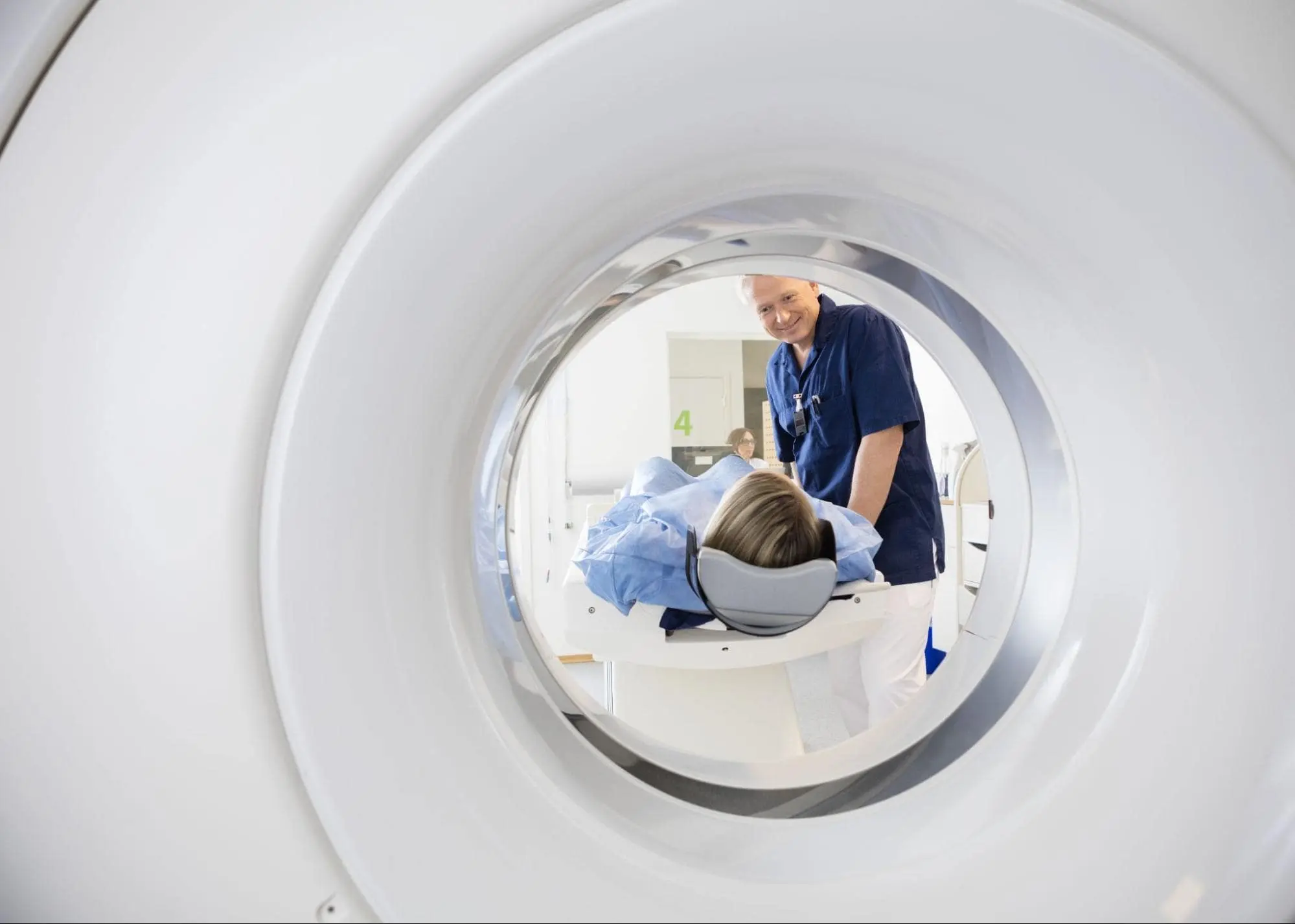
Do MRIs and X-rays Work?
MRI and X-ray imaging rely on different technologies. MRI uses powerful magnetic fields and radio waves to produce detailed images of the body’s internal structures. It offers excellent contrast resolution, making it easy to distinguish soft tissues, such as muscles, ligaments, and organs.
X-rays use electromagnetic radiation to create images based on tissue density. This makes X-rays very effective for viewing bones and other dense structures. They are less capable of capturing detailed images of soft tissue. X-ray images are usually single-plane, which can limit their usefulness for examining complex areas of the body.
What Works for Soft Tissue?
One of the primary benefits of MRI lies in its advanced soft tissue contrast. The ability to differentiate between various types of soft tissues helps detect subtle abnormalities, such as tears in ligaments or muscle injuries. When diagnosing nerve damage or the location of a tumor, this technology provides a level of detail that is not possible with X-ray imaging. This higher diagnostic accuracy may enhance the detection and evaluation of conditions like neurological disorders, muscle injuries, and soft tissue tumors.
What Are the Safety Features?
Safety is another area where MRI demonstrates advantages. Since MRI uses non-ionizing radiation, it eliminates the risks associated with exposure to ionizing radiation. This makes MRI particularly suitable for patients who require multiple scans over time. This includes those with chronic conditions or recurring injuries.
How Does Multi-planar Imaging Work?
Another significant strength of this technology lies in its ability to provide multi-planar imaging. Without repositioning the patient, this approach generates detailed anatomical images across different planes. This multi-layered perspective provides a comprehensive view of complex anatomical structures. It may be beneficial in areas such as the brain, joints, and spinal cord.
X-rays are limited to single-plane imaging. While this approach is sufficient for confirming fractures or checking for structural abnormalities in dense tissues, soft tissue injuries may lack the detail required for an accurate diagnosis. The lack of depth and perspective can hinder the ability to fully assess conditions affecting muscles, tendons, or nerves.
What Are Other Practical Applications?
MRI’s versatility extends across a range of specialties, from orthopedics to neurology, where soft tissue imaging plays a key role. MRI excels at providing comprehensive diagnostic solutions. Healthcare providers may rely on these detailed scans to formulate effective treatment plans tailored to each patient’s specific needs.
Find a Radiology Clinic for an MRI
The choice between MRI and X-ray ultimately depends on the specific diagnostic requirements. While X-ray remains ideal for imaging dense structures, such as bones, it falls short when detailed soft tissue imaging is required. With its superior contrast resolution, multi-planar imaging capabilities, and non-ionizing technology, MRI is better equipped to uncover the complexities of soft tissue anatomy and pathology. If you’re navigating options for soft tissue imaging, schedule an appointment with a radiologist near you.
- Where to Find South Slope Brooklyn News Online?
- How to Stay Updated with GlaadVoice.com
- How Do I Search for Manga on Manga Pill? A Beginner’s Complete Guide
- How Does OneFramework Improve Web Development? A Complete Beginner’s Guide
- Does GlaadVoice.com Cover Technology News?
Dr. Emma Green is a health and wellness expert with over 10 years of experience in nutrition and fitness. Passionate about helping others live their healthiest lives, Dr. Green shares practical advice on wellness, nutrition, and sustainable living through LivingSpristine.