Nuclear Stress Testing: The Key to Accurate Heart Disease Diagnosis
Heart disease remains a leading cause of health challenges worldwide. Timely and accurate diagnosis plays a key role in managing heart conditions effectively. Among the diagnostic tools available, nuclear stress testing is widely regarded as one of the most reliable methods. It aids in the assessment of heart function and blood flow. Here is more information on nuclear stress testing, its purpose, and how it works:
What is Nuclear Stress Testing?
A nuclear stress test is an advanced imaging procedure that evaluates blood flow to the heart, both during rest and when the heart is under stress. By using a small amount of radioactive material and specialized imaging equipment, medical professionals can create detailed pictures of how well blood moves through the heart’s chambers and arteries. This helps guide diagnosis and treatment protocols later on.
During the test, a patient undergoes two phases of imaging. The first phase involves capturing images of the heart while at rest. The second phase assesses blood flow during physical exertion, which can also be simulated with the use of medication. This radioactive material highlights areas with reduced or blocked blood flow, helping to pinpoint potential issues in the heart.
Why Do Doctors Use This Test?
This testing method helps identify problems that might remain unnoticed during other types of examinations. It is beneficial for detecting coronary artery disease (CAD), a condition where the arteries that supply blood to the heart become narrowed or blocked. It can also measure the heart’s pumping function and identify areas of the heart muscle that have been damaged from prior issues, such as a heart attack. These stress tests are valuable for gauging the effectiveness of current treatments by checking how well blood flow has been restored.
What Happens During the Procedure?
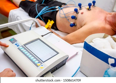
The process typically starts with an injection of a small, safe amount of radioactive material, called a radiotracer, into the bloodstream. This substance circulates through the blood and emits signals detectable by a special camera. Here is how the rest of the procedure typically goes:
- Imaging at Rest: Initial images of the heart are taken while the patient is at rest. These images act as a baseline against which stressed conditions can be compared.
- Creating Stress Conditions: The “stress” part of the test comes next. This is typically done in two ways. Physical exercise, such as walking on a treadmill or pedaling a stationary bicycle. If physical exercise isn’t suitable, medications are used to increase blood flow and mimic the effects of exercise on the heart.
- Repeat Imaging: After the heart has been “stressed,” another set of images is taken. A comparison of the resting and post-stress images allows healthcare providers to identify any areas with reduced blood flow or other abnormalities.
The procedure is non-invasive, with only minor discomfort from the injection or physical exertion.
Find a Cardiologist
Nuclear stress testing provides a detailed picture of heart health, enabling medical providers to make informed decisions about further care. By identifying areas of concern with precision, this test provides a valuable tool for enhancing overall heart care management. If you’re exploring diagnostic options for evaluating heart function, speak with a healthcare professional. They can help determine whether this type of stress testing is appropriate for your specific situation.
- What to Expect When Visiting a Foot and Ankle Specialist
- Causes of PTSD
- The Link Between Plantar Fasciitis and Weight Gain: What You Need to Know
- How Pet Ownership Can Positively Impact Life with Fibromyalgia
- The Importance of Stretching and Flexibility in Sports Medicine
Dr. Emma Green is a health and wellness expert with over 10 years of experience in nutrition and fitness. Passionate about helping others live their healthiest lives, Dr. Green shares practical advice on wellness, nutrition, and sustainable living through LivingSpristine.